NTNU’s NanoLab facility uses NanoSight NTA system to characterize nanoparticles.

NanoSight, leading manufacturers of unique nanoparticle characterization technology, reports on the characterization of nanoparticles at the NanoLab of the Norwegian University of Science & Technology, NTNU.

NTNU NanoLab is a cross faculty, strategic initiative with the objective to coordinate and promote nanoscience and nanotechnology at NTNU. The laboratories are well-equipped with state of the art instrumentation designed to be used by as many researchers from as many disciplines as possible.

For example, Katarzyna Psonka- Antonczyk is a post-doctoral fellow in the Biophysics and Medical Technology group within the Department of Physics. Her interests include the characterization of nanovesicles (exosomes) with sizes ranging from 30 to a few hundred nanometers secreted by cancer cells to the extracellular matrix. Exosomes are intercellular shuttle vehicles of various materials and contain information that can reprogram targeted cells. Exosomes contain membrane proteins, cytosolic proteins and small RNAs (miRNA). These vesicles are transported by bodily fluids (blood) and can likely fuse back with plasma membranes, introducing new proteins and RNA in new cells distant from the cell of origin. She is applying single-molecule techniques like atomic force microscopy and total internal reflection fluorescence microscopy to visualize individual exosomes and to characterize their membrane repertoire.

Knowing the concentration of secreted exosomes can facilitate estimation of the secretory abilities of cells and can help in further sample preparation. When coupled with fluorescent light, the NanoSight Nanoparticle Tracking Analysis system enables the analysis of exosome samples to provide information of various distinct subpopulations of vesicles by labeling with specific antibodies tagged with a fluorescent reporter. In the opinion of Dr Psonka-Antonczyk, the NanoSight LM10 has proven to be a very suitable instrument to access the concentration and size profile of exosomes.

Prior to using NanoSight, Dr Psonka-Antonczyk tried to employ dynamic light scattering but the results were rather irreproducible and not very reliable. In contrast, NanoSight exceeded her expectations. She said “NanoSight’s system is simple and easy to operate in providing information on the exosomes concentration and size profile in a very short time. I can also use it as a test measurement providing the first glance on the exosomes before running more elaborate and time-consuming experiments.”
The range of applications for NTA at NanoLab is diverse. For example, metallic and magnetic nanoparticles (Chemical Engineering) used for biomedical applications including targeted drug delivery and MRI contrast enhancement are studied by Dr Gurvinder Singh. He likes the NTA approach because “it provides the determination of particle concentration, better resolution of particle size and size distribution with real time visualization. This instrument is more impressive than DLS.”

Calorimetry As A Key Technique For Gas Adsorption Investigation

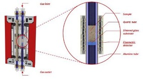
The investigation of gas adsorption on catalysts and solid sorbents depend upon a good interaction between the reactive gas and the powder. The SENSYS evo DSC is equipped with a crossing furnace, which allows the use of a quartz tube reactor. This makes it possible to operate in true plug-flow fixed bed reactor mode, widely used in heterogeneous catalysis, and thus precisely simulate industrial processes.

Additionally the exhaust gases can be flowed to an on-line gas analyzer such as a mass spectrometer, TCD or FTIR analyzer.

More complex thermal and gas flow profiles can provide adsorption / desorption / temperature programmed desorption data from a single experiment.

Example of application

  • Determination of the heat of adsorption / desorption of NH3 on different solid media. Simultaneous determination of the amounts of NH3 adsorbed / desorbed with coupled quantification techniques.
  • Catalytic oxidation of propane

Evaluation of the performance of catalysts for the selective catalytic reduction (SCR) of NO with NH3 and O2

See more in the following Aplication Note AN674 -Gas adsorption on catalysts and adsorbents using a quartz tube reactor on a Calvet type DSC

calorimetry

Witec Workshop – May 24th, 2013

WITec 3D Raman Imaging Meets AFM, SNOM and Profilometry Workshop. May 24th, 2013 – Université de Montréal

This workshop will give a detailed introduction to the operational principles and instrumental configurations relevant to confocal Raman and Scanning Probe Microscopy (AFM, SNOM, Pulsed Force Mode). Our Speakers will cover several aspects of Raman and SPM imaging and their fields of applications. An extensive system demonstration gives the participants the opportunity to see this knowledge applied using an alpha300 RSA (combined AFM & SNOM Confocal Raman Microscope).

Scientists interested in how to chemically identify and image the compounds of a sample at the highest spatial resolution are invited to participate. Typical research fields for confocal Raman imaging are nanotechnology, pharmaceutics and cosmetics, materials and polymer sciences, archaeology and geoscience, forensics, coatings, thin films and all fields where a clear identification of the distribution of chemical compounds is a necessity.

Program:

Friday, 24th May 2013
09.30 Welcome
09.45 Wei Liu – Introduction to Confocal Raman Imaging
10.15 Sylvain Cloutier – Hybrid Nanocrystal-based Heterostructures for Opto-Electronis
10.45 Klaus Weishaupt – 3D Raman Imaging Meets AFM, SNOM and Profilometry
11.30 Jean-Francois Masson – Characterization of photonics nanomaterials for plasmonic sensors
12.00 Lunch & Equipment Demonstration

The workshop is free of charge but it requires registration due to limited availability of places. In order to register, please contact Murielle Johnson at muriellej@aca.ca or 905-502-2088

Click here and download the May 24th 3D Raman Imaging Meets AFM, SNOM and Profilometry Workshop PDF for full program details.

Introducing the New Environmental Scanner for the Cypher™ AFM – the Highest Resolution Fast Scanning AFM

Imagine an AFM that routinely images the DNA double helix (top image) and atomic point defects (bottom) – in any environment.

We invite you to Booth 510 at the MRS Fall Meeting for an exclusive preview of the new Environmental Scanner for Cypher.

The Cypher Environmental Scanner is a modular and customizable platform with sample temperature control in a wide spectrum of environments. The Environmental Scanner is designed around a sealed cell which can sustain moderate positive pressures and uses chemically inert materials, such as fused silica and FFKM, to provide a controlled environment while maintaining accurate and distortion free imaging.

Cypher offers the widest range of scanning environments:

  • Buffers, solvents, inert gases, strong acids/bases
  • Liquid/gas perfusion
  • Continuous liquid flow
  • Temperature control from 0°C to 250°C
  • Optional electrical feedthroughs

If your experiments require a glove box, Cypher can also be operated in this configuration.

MFP-3D™ AFM – High Resolution,
Closed-loop Precision, and Flexibility

We’ll also be featuring the MFP-3D, the most versatile AFM platform with numerous environmental accessories and advanced scanning modes. The MFP-3D can be configured as:

Both the MFP-3D and Cypher AFMs scan with all standard AFM imaging modes (tapping, contact, MFM, phase, etc.), and numerous exclusive imaging modes:

SUMMER SCHOOL AND WORKSHOP IN CALORIMETRY 2015 CALORIMETRY AND THERMAL METHODS IN MATERIAL SCIENCE

LYON, FRANCE – JUNE 21-26, 2015

Calorimetry and thermal analysis methods, alone or linked to other techniques, will be described and applied to the characterization of catalysts, oxides, metals, supports, adsorbents, polymers, composites, food, pharmaceuticals, mesoporous solids, ionic liquids, phase change materials, heat storage materials, hydrogen storage materials. Examples will be given in various domains: catalytic reactions, air and wastewater treatment, clean and renewable energies, refining of hydrocarbons, green chemistry, hydrogen production and storage, CO2 capture, interseasonal heat storage, food chemistry, polymers…

Making high performance affordable

Atomic force microscopes (AFM) offer atomic-scale resolution for imaging applications in life sciences and materials research.

But AFM’s often come with a very expensive price tag – which can make it a challenge for scientists to afford this technology in these days of shrinking research budgets.

Earlier this month, our partners at Asylum Research introduced a high performance solution that expands the reach of atomic force microscopes to researchers with limited budgets for imaging instrumentation. This new AFM eliminates some of the more advanced accessories and features of the high-end models without sacrificing core performance.

“This is in stark contrast to some competitors who have taken old technology and repackaged it as entry-level AFMs with lower performance,” says Ben Ohler, AFM Business Manager, in an Asylum Research news release. “(This model) is based on the technology-leading MFP-3D AFM, known for closed-loop precision, high-resolution imaging, and low-noise force measurements.”

This model – the MFP-3D Origin – is great news for the AFM research community. It offers far more than just the basic scan modes that you’d expect in a more affordable model and it includes many advanced modes such as nanolithography, Dual AC Mode and piezoresponse force microscopy.

Like the higher-performance model it’s based on, it can be used for a variety of applications in material science, physics, data storage and semiconductors, polymers, chemistry, biomaterials and bioscience. The MFP-3D can be used for single molecule mechanical experiments on DNA, protein unfolding and polymer elasticity, as well as force measurements on biomaterials and polymers.

Until now, purchasing a lower cost AFM meant sacrificing resolution, scan speed, or the availability of sophisticated imaging modes. With the advent of the Origin from Asylum Research, a limited budget does not translate into limited research.

Please contact us and speak to one of our sales representatives to request a quote or more information on any of our products.